PREVALENCE OF HELICOBACTER PYLORI AMONG STUDENTS OF DR. JOHN ADAH COLLEGE OF HEALTH TECHNOLOGY OTUKPO, BENUE STATE NIGERIA.
BY
ODOABUCHI TITUS UCHECHUKWU
MATRIC NO: JASHT/MLT/2013/026
BEING A PROJECT WORK PRESENTED TO THE DEPARTMENT OF MEDICAL LABORATORY SCIENCE. DR. JOHN ADAH COLLEGE OF HEALTH TECHNOLOGY, OTUKPO, BENUE STATE.
IN PARTIAL FULFILMENT OF THE REQUIREMENT FOR THE AWARD OF DIPLOMA IN MEDICAL LABORATORY SCIENCE.
CERTIFICATION
This is to certify that this project work was carried out by Odoabuchi Titus Uchechukwu of Department of medical laboratory science, Dr. John Adah College of Health Technology Otukpo, Benue State. Under the supervision of MLS Kelvin Anayo.
ACKNOWLEDGMENT
With the deepest gratitude, I wish to thank the Almighty God for His love, mercy, wisdom, knowledge and understanding He had given to me and towards this documentation; all thanks goes to Him.
I would like to acknowledge and express my gratitude to the following people for their magnificent support and contribution to my journey of life: my parents Evan. Odoabuchi Peter and Mrs. Odoabuchi Serah, my siblings and all members of my family for their financial, moral and academic encouragement to make my dream a reality.
I am grateful to my HOD MLS. Kelvin Anayo for the intellectual contribution and his advice towards my academic pursuit. May God give you long life.
Gratitude to my classmates and all my friends most especially to Ogenyi Michael for their advice, may God Almighty continue to bless and guid you all in anywhere you find your selves in Jesus name (Amen)
DEDICATION
This project work is been dedicated to God Almighty who gave me the wisdom, knowledge and the understanding to make this project paper to be a reality, I say may his name alone be praise.
I also want to dedicate this project work to my parents whom God has used as a source of support to me, spiritually, materially and financial. I say may God strengthen you people the more in Jesus Name (Amen).
ABSTRACT
Helicobacter pylori is a gram negative spiral bacterium that causes gastritis and pyloric ulcers. The aim of the study was to determine the prevalence of Helicobacter pylori infection among the students of Dr. John Adah College of Health Technology. The Objectives of study was to evaluate the level of knowledge the student have about the disease and to access the compliers of student to the sanitary measure toward eradicating the disease. Transmission of the infections is thought to occur as a consequence of direct human-to-human transmission via either the oral-oral route or perhaps the fecal-oral route or both. The bacteria is found commonly in the lining of the stomach and the duodenum because they adapt well to the acidic low PH environment. The methodology adopted was experimental design, while blood sample was collected and analyzed. The population used at the course of the study was one hundred samples. The information obtained was represented using frequency table. After the analysis, it is clearly defined that the prevalence of Helicobacter pylori infection among the student is very high with infectivity rate of 57%. This is because most of the students does not have knowledge about the disease and also they are not practicing adequate personal hygiene.
CHAPTER ONE
INTRODUCTION
Helicobacter pylori, previously called campylobacter pylori are a gram negative, microaerophilic bacterium found usually in the stomach. It is also describe as a helix-shaped (classified as a curved rod, not spirochaete) gram-negative bacterium about 3µm long with a diameter of about 0.5µm. it is microaerophilic because it requires oxygen, but at lower concentration than is found in the atmosphere. It contains a hydrogenise which can be used to obtain energy by oxidizing molecular hydrogen (H2) produced by intestinal bacteria.
The bacteria infects or gets into the body through contaminated food or water with the bacteria. Since it is present in human saliva, it can spread through mouth-to-mouth contact, such as kissing.
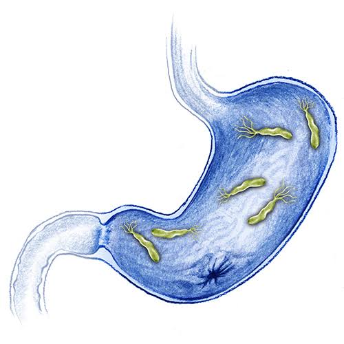
After H. Pylori enters the body, to avoid the acidic environment of the interior of the stomach (lumen), Helicobacter Pylori uses it’s flagella to burrow into the mucus lining of the stomach to reach the epithelia cells underneath, where the PH is more neutral. Helicobacter Pylori have the ability to sense the PH gradient in the mucus membrane and move towards the less acidic region (chemotaxis). This also keeps the bacteria from being swept away into the lumen with the bacteria’s mucus environment, which is constantly moving from its site of creation at the epithelium to its dissolution at the lumen interface. It lives in the mucus that coats the lining of the stomach and duodenum and produces urease, an enzyme that neutralizes stomach acid by making it less acidic. It adheres to the epithelial cells by producing adhesions which bind to lipids and carbohydrates in the epithelial cell membrane. It attacks the lining of the stomach, which usually protects the body from the acid the body uses to digest food. Once the bacteria have done enough damage, acid can get through the lining, which leads to ulcers or gastritis (inflammation).
These may bleed, cause infections, or keep food from moving through the digestive tract.
In addition to using chemotaxis to avoid areas of low PH, Helicobacter Pylori also neutralize the acid in its environment by producing large amounts of urease, which breaks down the urea present in the stomach to carbon dioxide and ammonia. The ammonia, which is basic, then neutralizes stomach acid.
Helicobacter Pylori were first discovered in the stomach of patients with gastritis and ulcers in 1982 by Dr. Barry Marshall and Robin Warren of Perth, Australia. At that time, the conventional thinking was that no bacterium could live in an acid environment of the human stomach. In recognition of their discovery Marshall and Warren were awarded the 2005 Nobel Prize in physiology or medicine.
Before the research of Marshall and Warren, German scientists found spiral-shaped bacteria in the lining of the human stomach in 1875, but they were unable to culture them, and the results were eventually forgotten. The Italian researcher Giulio Bizzozero described similarly shaped bacteria living in the acidic environment of the stomach of dogs in 1893.
Professor Walery Jaworski of the Jagiellonian University in Krakow investigated sediments of gastric washing obtained by lavage from human in 1899. Among some rod-like bacteria, he also found bacteria with a characteristic spiral shape, which he called Vibrio rugula. He was the first to suggest a possible role of this organism in the pathogenesis of gastric disease. Several small studies conducted in the early 20th century demonstrated the presence of curved rods in the stomachs of many people with peptic ulcers and stomach cancers.
Interest in understanding the role of bacteria in stomach disease was rekindled in the 1970s, with the visualization of bacteria in the stomachs of people with gastric ulcers.
The bacteria had also been observed in 1979, by Robin Warren, who researched it, further with Barry Marshall from 1981. After unsuccessful attempts at culturing the bacteria from the stomach, they finally succeeded in visualization colonies in 1982, when they unintentionally left their petri dishes incubating for five days over the Easter weekend. In their original paper, Warren and Marshall contended that most stomach ulcers and gastritis were caused by bacterial infection and not by stress or spicy food, as had been assumed before. To demonstrate H. Pylori caused gastritis and was not merely a bystander, Marshall drank a beaker of H. Pylori culture. He becomes ill with nausea and vomiting several days later. An endoscopy 10days after inoculation revealed signs of gastritis and the presence of H. Pylori. These results suggested Helicobacter Pylori was the causative agent.
In 1994 the National Institutes of Health stated most recurrent duodenal and gastric ulcers were caused by H. Pylori and recommended antibiotics are included in the treatment regimen.
The bacterium was initially named campylobacter pyloridis, then renamed C. Pylori. In 1989 other research showed that the bacterium did not belong in the genus campylobacter, it was placed in its own genus Helicobacter.
Up to 85% of people infected with Helicobacter Pylori never experience symptoms or complication. Acute infection may appear as an acute gastritis with abdominal pain (stomach ache) or nausea. Where this develops into chronic gastritis, the symptoms, if present, are often those of non-ulcer dyspepsia: stomach pains, nausea, bloating, belching and sometimes vomiting or black stool.
However, with a good health habits, someone can protect him or herself from Helicobacter Pylori. It is more common in countries or communities that lack clean water or good sewage system.
AIM OF STUDY
The aim of this study was to determine the level of endemicity of Helicobacteriosis among student attending Dr. John Adah College of Health Technology Otukpo.
OBJECTIVE OF STUDY
To identify the complication and risk factor that aids the transmission of the infection
To evaluate the level of knowledge the students have about the disease
To access the compliers of student to the sanitary measure toward eradicating the infection.
CHAPTER TWO
2.0 LITERATURE REVIEW
2.1 DEFINITION OF HELICOBACTER PYLORI
Helicobacter pylori is a gram negative spiral bacterium that causes gastritis and pyloric ulcers in humans. The bacterium has also been linked with other gastrointestinal diseases including gastric cancer and gastric mucosa-associated lymphoid tissue (MALT) lymphonia.
Helicobacter Pylori is found commonly in the lining of the stomach and the duodenum because they adapt well to the acidic, low PH environment. Urease is the central metabolism of Helicobacter pylori. In order to survive, the organism uses urea to produce ammonia and bicarbonate to neutralize the acid in the stomach. The metabolic products from Helicobacter pylori can alter the host, and change the acidity of the environment and increase the supplements of nutrient to colonize the stomach.
J Ochei, Medical laboratory science Theory and practice (page: 711) describe that Helicobacter pylori is a small “S” shaped gram negative bacteria. They are microaerophilic, catalase and oxidase, rapidly urease positive. Helicobacter organisms were originally placed in the genus campylobacter. Phylogenic studies have shown that they are not related to the group and have been classified as a new genus Helicobacter.
Monica Cheesbrough, District Laboratory Practice in tropical countries part 2 (page: 197) explain that Helicobacter pylori is formerly called campylobacter pylori which spread widely in developing countries, 8 in 10 children by age 5years and more than 90% of adults are infected. Transmission is by person to person contact, and probably also by contaminated water and food.
In most person, infection with Helicobacter pylori is asymptomatic. In others, colonization of the internal surface lining of the stomach wall causes inflammation and chronic gastritis which predispose to ulceration. Helicobacter pylori is thought to be the causes of most gastric and duodenal ulcers.
According to pocket medical Dictionary (page 139), Helicobacter pylori is a bacterium causing a number of gastrointestinal diseases via gastric infection. These include peptic ulceration, gastric cancer and MALToma.
Farlex partner medical Dictionary defined Helicobacter pylori as a bacterial species that produces urease and causes gastritis and nearly all peptic ulcer disease of the stomach and duodenum. Infection with this organism also plays an etiologic role (probably along with dietary co-factors) in dysplasia and metaplasia of gastric mucosa, distal gastric adenocarcinoma, and non-Hodgkin lymphoma of the stomach. They are curved or spiral, flagellated gram-negative bacillus, Helicobacter pylori colonize the gastric mucosa, attaching itself to the surface of mucus-secreting columnar cells. The ability of the organism to survive in an acid medium is due to its production of urease, which converts urea to ammonia and alkalinizes the film of mucus in which it resides. Originally believed to be a species of campylobacter, the organism was reclassified as Helicobacter pylori in 1989. Transmission is believed to be from person-to-person by the fecal-oral route.
2.2 SCIENTIFIC CLASSIFICATION OF HELICOBACTER PYLORI
Higher order taxa
Domain: Bacteria
Phylum: Proteobacteria
Class: Epsilonproteobacteria
Order: Campylobacterales
Family: Helicobacteraceae
Genus: Helicobacter
Species: Helicobacter Pylori
2.3 MORPHOLOGY OF HELICOBACTER PYLORI
Helocobacter pylori is a gram-negative bacterium, measuring 2 to 4µm in length and 0.5 to 1µm in width. Although usually spiral-shaped, the bacterium can appear as a rod, while coccoid shapes appear after prolonged in vitro culture or antibiotic treatment. These coccoids cannot be cultured in vitro and are thought to represent dead cells, although it has been suggested that coccoid forms may represent a viable, non-culturable state. The organism has 2 to 6 unipolar sheathed flagella of approximately 3µm in length, which often carries a distinctive bulb at the end. The flagella confer motility and allow rapid movement in viscous solutions such as the mucus layer over lying the gastric epithelia cells. In contrast to many other pathogens of the gastrointestinal tract, it lacks fimbrial adhesions.
2.4 GROWTH REQUIREMENT OF HELICOBACTER PYLORI
A key feature of Helicobacter pylori is its microaerophilicity, with optimal growth at O2 level of 2 to 5% and the additional need of 5 to 10% Co2 and high humidity. There is no need for H2, although it is not detrimental to growth. Many laboratories utilize standard microaerobic conditions of 85% N2, 10% Co2 and 5% O2 for Helicobacter pylori culture. Growth occurs at 34 to 4OOc, with an optimum of 37OC.
Although its natural habitat is the acidic gastric mucosa, Helicobacter pylori is considered to be a neutrolophile. The bacterium will survive brief exposure to PH of <4, but growth occurs only at the relatively narrow PH range of 5.5 to 8.0, with optinal growth at neutral PH.
Helicobacter pylori is fastidious micro-organism and requires complex growth media. Often these media are supplemented with blood or serum. These supplements may act as additional sources of nutrients and possibly also protect against the toxic effects of long-chain fatty acids. Commonly used solid media for routine isolation and culture of Helicobacter pylori consist of Columbia or brucella agar supplemented with either (lysed) horse or sheep blood or alternatively, newborn or fetal calf serum. Liquid media usually consist of either brucella, Mueller-Hinton, or brain heart infusion both supplemented with 2 to 10% calf serum, often together with either dent or skirrow supplement
2.5 EPIDEMIOLOGY
The prevalence of Helicobacter pylori shows large geographical variations. Approximately 50% of the world’s population has been estimated to be infected. In various developing countries, more than 80% of the population is Helicobacter pylori positive, even at young age. The prevalence of Helicobacter, pylori infection in industrialized countries generally remains under 40% and is considerably lower in children and adolescents than in adults and elderly people. Within geographical areas, the prevalence of Helicobacter pylori inversely correlates with socioeconomic status, in particular in relation to living conditions during childhood. However, the infection rate of children in developing nations is higher than in industrialized (developed) nations probably due to poor sanitary conditions, perhaps combined with lower antibiotics usage for unrelated pathologies. In developed nation, it is currently uncommon to find infected children, but the percentage of infect people increase with age, with about 50%infected for those over the age of 60 compared with around 10% between 18 and 30 years. This higher prevalence among the elderly reflects higher infection rates in the past when the individuals were children rather than more recent infection at a later age of the individual. The active elimination of Helicobacter pylori from the population and improved hygiene and housing conditions have resulted in a lower infection rate in children, which is reflected in the age distribution of this lifelong colonizing bacterium. Overall, new infection more commonly occurs in childhood and lasts for life unless specifically treated.
2.6 PATHOGENESIS OF HELICOBACTER PYLORI
These remarkable organisms survive the extreme acidity of the stomach because of their powerful urease. This enzyme creates an alkaline environments by hydroxyzine urea to ammonia and carbon dioxide. Urea is a waste product of protein catabolism by the body’s cells and is normally present in the gastric juices.
Once the bacteria reach the mucus that coats the stomach or duodenal lining, they use their flagella to burrow into the mucus lining of the stomach to reach the epithelial cells. In this location the PH of the mucus is nearly neutral, and the bacteria attach to the mucus-secreting epithelium. The bacterial products incite an inflammatory response in the wall of the stomach, and mucus production decreases. Once infection occurs it persists for years, often for life it not treated.
The primary disorder, which occurs after colonization with Helicobacter pylori is an acute and chronic active gastritis. This condition can be observed in all Helicobacter pylori-positive subjects (individual). The intragstric distribution and severity of this chronic inflammatory process depend on a variety of factors, such as characteristics of the colonizing strain, host genetic and immune response, diet and the level of acid production. Helicobacter pylori-induced ulcer disease, gastric cancer and lymphoma, all these are complication of this chronic inflammation; ulcer disease and gastric cancer in particular occur in those individuals and at those sites with the most severe inflammation.
From 10% to 20% of infected persons develop ulcers; 60% to 80% of patients with gastric ulcers and 95% of those with duodenal ulcers are infected with Helicobacter pylori. The thinning of the protective mucus layer at the site of infection probably accounts for the development of peptic ulcers of the stomach and duodenum. A very small percentage of individuals infected with Helicobacter pylori develop cancer of the stomach, but more than 90% of those with stomach cancer are infected by the bacterium.
2.7 IMMUNE RESPONSE TO HELICOBACTER PYLORI INFECTION
Infection of the stomach with Helicobacter pylori induces an immune response with infiltration of the mucosa by macrophages, neutrophils and lymphocytes.
The immune response towards Helicobacter pylori can be divided into an innate and an adaptive response. During the infection both of them are activated. The innate response towards Helicobacter infection is generally an initial non-specific process, which reacts quickly with several bacterial molecules to signal infections danger and with the aim of killing the bacteria. By contrast, the adaptive immune response is delayed, antigen-specific, leads to the activation of T-cell, B-cell and memory cells and is shaped by the innate immune response.
Although the bacterium is rarely eliminated and infections can last for decades if left untreated. Helicobacter pylori has evolved several mechanisms to increase its adherence and persistence in the host. Elimination of Helicobacter pylori by phagocytes is inefficient because Helicobacter pylori exhibit several virulence factors to evade optimization, retard phagocytosis and disrupt membrane trafficking and phantom maturation after internalization of the micro-organism.
2.8 SIGNS AND SYMPTOMS OF HELICOBACTER PYLORI INFECTION
Most people with Helicobacter pylori do not have any symptoms. When the infection leads to an ulcer and other illness, symptoms may include abdominal pain, especially when the stomach is empty at night or of few hours after meals. The pain is usually described as a gnawing pain, and it may come and go. Eating or taking antacid drugs may relieve this pain.
A number of other symptoms which may be associated with Helicobacter pylori infection are:
Excessive burping
Feeling bloated
Nausea or vomiting
Lack of appetite
Unexplained weight loss
Belching
Passing dark or tarry like stool
Fatigue
Decreased appetite
Heartburn
Abdominal discomfort
Bad breath
2.9 COMPLICATION OF HELICOBACTER PYLORI INFECTION
Helicobacter pylori infections can lead to Gastritis (inflammation) and ulcers as earlier mention, but the infection or the ulcer itself can lead to more serious complications. These include:
Internal bleeding, which can happen when a peptic ulcer breaks through the blood vessel.
Obstruction, which can happen when an ulcer blocks food from leaving the stomach.
Perforation, which can happen when an ulcer breaks through the stomach wall.
Peritonitis, which is an infection of the peritoneum, or the lining of the abdominal cavity.
Studies show that infected people also have an increased risk of gastric adenocarcinoma, which is a type of stomach cancer.
2.10 RISK FACTOR OF HELICOBACTER PYLORI INFECTION
Almost about 90% of Nigerian populations are critically in high risk of helicobacter pylori infection. This is because the sources of this infection are too many of which man use them as their own source of food and water supply. Sources like streams either natural or artificial well, ponds and even pipe water supply this is because the organism can survive the conventional method of water treatment some time, also not all Nigerian practice adequate personal hygiene and as such faecal oral route as a result of poor environmental sanitation is another problem. Contaminated fruits when not washed properly can cause the infection. All vegetable which are fertilized by human/animals faeces when not well cooked can poses higher risk of this infection. The practice of pre-mastication is another risk of the infection to children, though they are much more at risk of this infection due to poor immunity on their part.
2.11 LABORATORY DIAGNOSIS OF HELICOBACTER PYLORI
Various tests have been developed for the detection of Helicobacter pylori, each with their specific advantages and disadvantages. The available tests are generally divided into invasive tests, based on gastric specimens for histology, culture or other methods, and noninvasive tests based on peripheral samples, such as blood, breath samples, stools, urine or saliva for detection of antibodies, bacterial antigens or urease activity. The choice of a specific test for an individual depends on local experience and the clinical setting. In research protocols, a combination of two methods is often applied. In daily clinical practice, use of a single test is generally adequate, and most tests are sufficiently accurate to be used for this purpose. For routine diagnostic purposes, histology, urea breath testing and culture are currently most often used, whereas the use of serology is most appropriate for large epidemiological studies. In hospital based care, many patients undergo endoscopy, which is then combined with an invasive test for Helicobacter pylori. Otherwise, breath test and serology are commonly used for children, faecal antigen tests offer the opportunity to assess Helicobacter pylori status without the need for endoscopy or vena puncture.
2.12 MANAGEMENT OF HELICOBACTER PYLORI INFECTION
Helicobacter pylori is managed or treat with certain antibiotics. However, a combination of medicines is needed to get rid of it completely. This is referred to as combination therapy although because it eradicates (gets rid of) the germ it is also referred to as eradication therapy. The patient needs to take two antibiotics at the same time. In addition, the patient needs to take a medicine to reduce the acid in the stomach. This allows the antibiotics to work well in the stomach. It is important to take all the medication exactly as directed and to take the full course.
Eradication therapy clears Helicobacter pylori in up to 9 in 10 cases if it is taken correctly for the full course. If you do not take the full course then the chance of clearing the infection is reduced. A second course of eradication therapy, using different antibiotics, will usually work if the first course does not clear the infection. Eradication therapy is sometimes called triple therapy as it involves three medicines; two antibiotics and an acid suppressing medicine.
Some of the drugs that are used in a triple therapy treatment include:
Clarithromycin
Metronidazole
Amoxicillin
Proton-pump inhibitors (PPI), such as pantoprazole, esomeprazole, or lansoprazole.
2.13 MODE OF TRANSMISSION OF HELICOBACTER PYLORI
Helicobacter pylori has a narrow host range and is found almost exclusively in humans and some non-human primates. Helicobacter pylori has on rare occasions been isolated from pet animals; thus, the presence of pets many be a risk factor for Helicobacter pylori infection. As conclusive evidence for zoonotic transmission of Helicobacter pylori is not yet available, new infections are thought to occur as a consequence of direct human-to-human transmission, via either the oral-oral route (through vomitus or possibly saliva by kissing or pre-mastication for children) or perhaps the fecal-oral route or both. Helicobacter pylori has been detected in saliva, vomitus, gastric refluxate and faeces, but there is no conclusive evidence for predominant transmission via any of these products. This may be due to the fact that most research on transmission has focused on them. The person-to-person mode of transmission is supported by the higher incidence of infection among institutionalized children and adults and the clustering of Helicobacter pylori infection within families.
Waterborne transmission, probably due to faecal contamination, may be an important source of infection, especially in part of the world in which untreated water is common. The most recent reservoir suggested for Helicobacter pylori transmission is the housefly.
However evidence is lacking those Helicobacter pylori can be transmitted to humans from flies that have been in contact with Helicobacter pylori infected faeces.
2.14 PREVENTION AND CONTROL OF HELICOBACTER PYLORI
Helicobacter pylori is a major cause of certain disease of the upper gastrointestinal tract rising antibiotic resistance increase the need to search for new therapeutic strategies, this might include prevention in form of vaccination. Much work has been done on developing viable vaccines aimed at providing an alternative strategy to control Helicobacter pylori infection and related disease, including stomach cancer.
Researchers are studying different adjuvant, antigens and routes of immunization to ascertain the most appropriate system of immune protection; however most of the research only recently moved from animal to human trials. Therefore, the only way now to control or prevent the infection is by the practice of personal hygiene which some of them include:
Wash hands properly after using the bathroom or toilet and also before preparing or eat food. Teach your children to do the same.
Avoid food or water that is not clean.
Do not eat any food that is not cooked thoroughly.
Avoid food served by people who have not washed their hands
CHAPTER THREE
3.0 RESEARCH METHODOLOGY
This chapter presents the methods, materials, reagent and procedure carried out to established objectives of this research work.
3.1 RESEARCH DESIGN
Experimental design was used to obtain information for this study. This study was designed to look into the prevalence of Helicobacter pylori among students attending Dr. John Adah College of Health Technology Otukpo Local Government Area of Benue State.
3.2 AREA OF STUDY
The area of my study is restricted to student attending Dr. John Adah College of Health Technology Otukpo Local Government Area of Benue State.
3.3 POPULATION OF THE STUDY
The target population of the study is 100 students of Dr. John Adah College of Health Technology Otukpo Local Government Area of Benue State.
3.4 SAMPLE AND SAMPLING TECHNIQUES
100 blood samples were collected randomly from the students of Dr. John Adah College of Health Technology. The blood sample was collected using EDTA anticoagulated container and labeled with the student name and other information needed. The samples were analysed at Dr. John Adah College of Health Technology Teaching Laboratory.
3.5 MATERIALS USE FOR SAMPLE COLLECTION & EXAMINATION
The materials include:
EDTA anticoagulated container
Sterile needle and syring
Tourniquet
Cotton wool
Methylated spirit
Hand glove
Plastic pipettor
H. pylori rapid test cassette
3.6 METHOD OF BLOOD SAMPLE COLLECTION
Procedure for blood sample collection are:
A sterile, dry, preferably plastic syringe of the capacity required was selected.
A tourniquet was apply to the upper arm of the patient to enable the veins to be seen and felt.
The index finger was used to feel and select suitable vein that is sufficiently large, straight that does not roll and with a direction that can be felt.
The puncture site was cleaned with methylated spirit and allow to air dry.
The thumb of the left hand was used to hold down the skin below the puncture site.
The venipuncture was made with the bevel of the needle directed upwards in the line of the vein. The plunger of the syringe was withdrawn steadying to fill the syringe with required blood.
When sufficient blood has been collected, the tourniquet was release and the patient was instructed to open his first.
The needle was removed immediately from the puncture site and a piece of dry cotton wool was used to press on the puncture site.
The patient was instructed to continue pressing on the puncture site until the bleeding has stopped.
The needle was removed from the syringe and carefully filled the container with the required volume of blood. The needle was discarded safely.
The blood was mix immediately in the anticoagulated container.
All specimens were labeled with the student’s name, age, sex and the date of collection.
The samples were taken to the laboratory of Dr. John Adah College of Health Technology for immediate examination.
3.7 METHOD OF SAMPLE ANALYSIS
The method used is the H. Pylori Rapid Test Device for the diagnosis of Helicobacter pylori
3.7.1 TEST PROCEDURE
The test cassette was removed from the pouch
The test cassette was placed on clean and leveled bench
Using the plastic pipettor provided, one drop of whole blood was draw into the sample will of the cassette
One drop of butter was also added into the sample well
It was allowed to stand for 10minutes.
3.73 INTERPRETATION
Double lines (one on test region and the other on control region) indicate positive result.
While one line in the control region only indicate negative result. But if there is no colour band in both control and test line, it indicate invalid test result. This is an indication of a possible error in performing the test. The test should be repeated using a new device.
3.8 METHOD OF DATA ANALYSIS
The sample obtained from students of Dr. John Adah College of Health Technology was simply expressed in percentages for easy statistical understandings and were recorded in tabular forms.
CHAPTER FOUR
4.0 PRESENTATION OF DATA
In this research work, 100 blood samples were collected from student of Dr. John Adah College of Health Technology Otukpo. 50 samples were collected from male and 50 samples were collected from female. After the analysis, 57 samples were tested positive while 43 samples were tested negative. In respect to gender, 21 samples among the 50 samples collected from male were positive and 29 were negative, 36 samples among the 50 samples collected from female were positive and 14 were negative. The tables below shows more detail of the result.
4.1 Table 1: showing the infectivity rate of H. pylori among the students
No of sample
Positive
% pos
Negative
% Neg
100
57
57%
43
43%
4.2 Table 2: showing the prevalence of Helicobacter pylori infection among students in respect to gender.
Gender
No of pos sample
% pos
No. of Neg sample
% Neg
Male
21
37%
29
67%
Female
36
63%
14
33
Total
67
100%
43
100%
CHAPTER FIVE
5.0 DISCUSSION, CONCLUSION AND RECOMMENDATION
5.1 DISCUSSION OF FINDING
This research intended to establish the prevalence of Helicobacter pylori infection among student of Dr. John Adah College of Health Technology Otukpo. In this research all the people in Dr. John Adah College of Health Technology Otukpo constituted the population. Out of this population, one hundred samples were collected.
At the course of the study, it was discovered that there was high prevalence of Helicobacter pylori infection among the students with 57% infectivity rate among the 100 sample analyzed. The research results confirm the previous research of Collins which said that half of the world population are infected with Helicobacter pylori. The result obtained from the research showed that the student are not complying to sanitary measure toward eradicating the infection (H.Pylori), that is, they are not practicing adequate personal hygiene. According to the finding of the research work, it showed that the prevalence rate is higher on the female part than the male.
5.2 CONCLUSION
From the finding of this study, it is clearly defined that the prevalence of Helicobacter pylori infection among students of Dr. John Adah College of Health Technology Otukpo was very high. This is because most of the students does not have knowledge about the disease and also they are not practicing adequate personal hygiene. This study highlighted the importance of practicing adequate personal hygiene and proper disposal and faecal management as these are keys for the prevention of this infection.
5.3 RECOMMENDATION
Based on the findings that have been made from students attending Dr. John Adah College of Health Technology Otukpo, the researcher has come with the following suggestions and recommendation.
Health education should be given to the student on how to practice adequate personal hygiene.
Those that are infected with the bacteria should be treated with appropriate medication.
Health education should also be given to the student on the proper disposal and faecal management, these are keys for the prevention of this infection.
Further studies should be carried out for more investigation in this institution.